Examples of QEEG Brain Maps
These are examples of brain maps and a variety of disorders. These examples are not intended to be representative of each type of disorder.
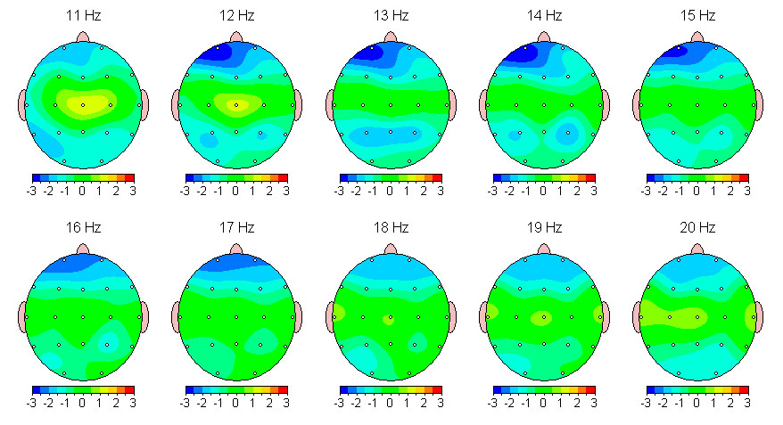
Depression
This is a brain map from a male in his 20's with severe depression that did not respond to medication. The map shows deficient beta in the left prefrontal cortex. This is consistent with research that shows hypoperfusion (decreased blood flow) in the this region of the brain in depression. This client was treated with neurofeedback on the areas of the center and left forehead. Decreased depression was reported within the first several sessions. The depression has been in complete remission for more than 2 years.
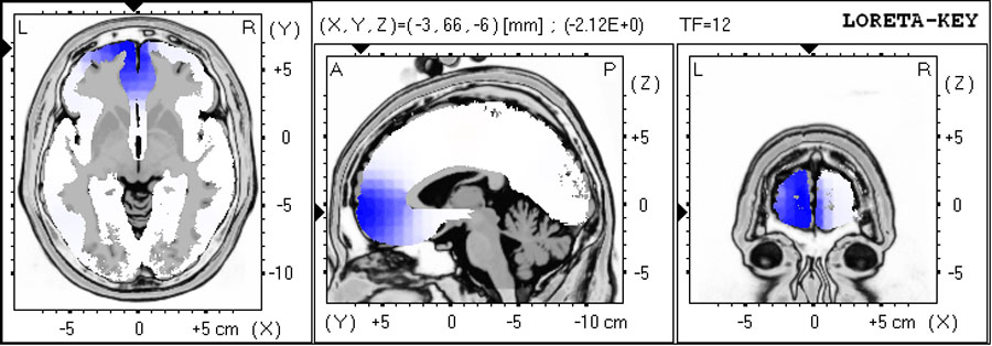

Obsessive Compulsive Disorder
Over-active posterior cingulate shown in LORETA analysis.


Attention Deficity Disorder (ADHD): Excess Theta Type
These quantitative EEG maps show the excess theta type of ADHD with excess 6 Hz in the right frontal area. There is also problems with depression (left front) and learning disability (back of head).

Attention Deficity Disorder (ADHD): Excess Beta Type
These maps show excess beta EEG activity.

Bipolar Disorder (Unstable)

Combat related PTSD
Severe depression, nightmares, anxiety, anger.


